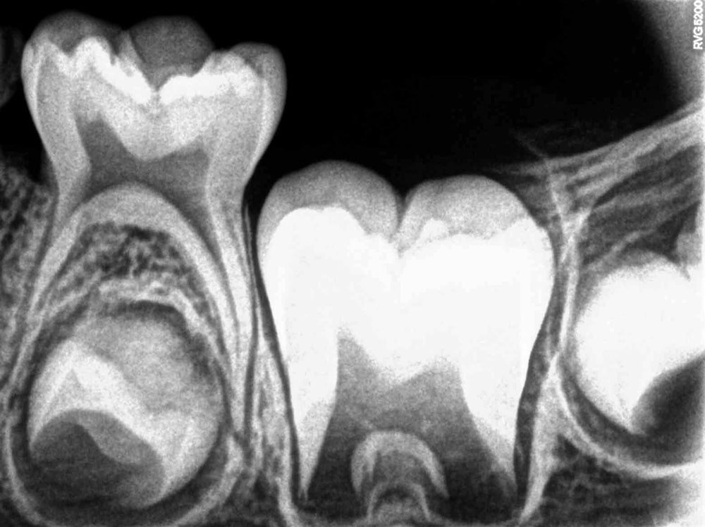
Radiovisiography (RVG)
RVG is one of the latest imaging technologies in dentistry used for routine dental examination with minimal radiation and it covers only a few teeth and offers a two-dimensional evaluation of the offending tooth.
Unlike the conventional film radiography, it is acquired with reduced radiation to the patient and its software gives clinicians the ability to enlarge the different areas of the image for better diagnosis. Undoubtedly, the competence of the Carestream RVG software to achieve radiographs with no loss of image quality and ability to archive for longer periods of time, make the use of conventional dental film radiographs obsolete.
CBCT is a specialized type of X-ray imaging technology that produces three-dimensional images of the area being scanned.It provides high-resolution images, allowing detailed visualization of dental structures and tissues. The radiation is of lower dose compared to traditional CT scans, making it safer for patients.
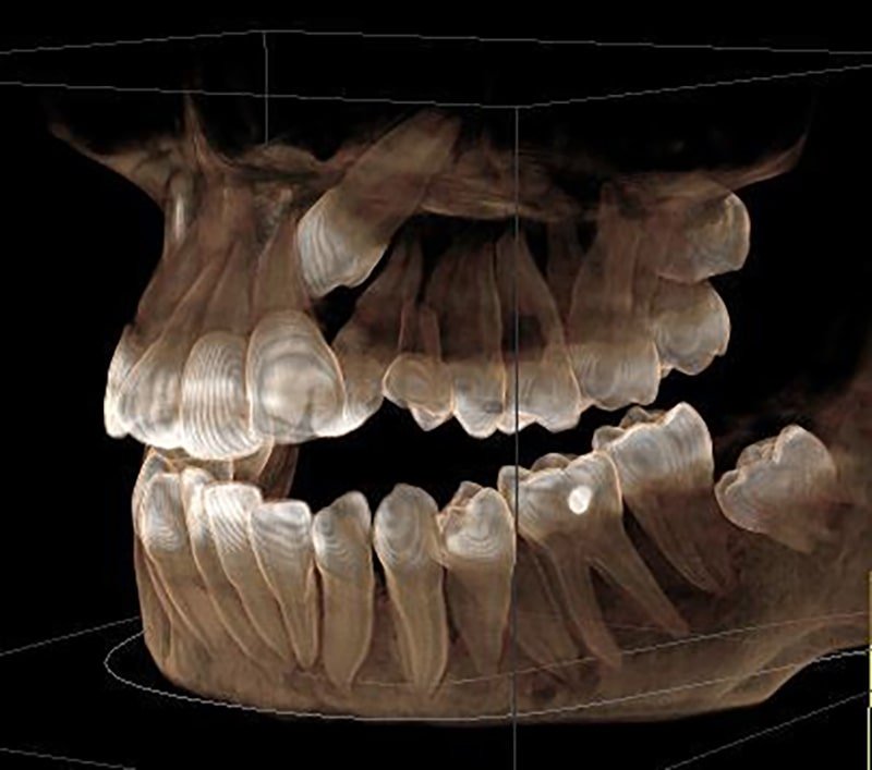
CBCT is a very important tool in assessing the bone quality and quantity, which is necessary for dental and maxillofacial procedures. It can also visualize the soft tissues, such as nerves and blood vessels and their path, aiding in precise digital treatment planning and thus avoiding any damage to the vital structures or any complications. It allows for precise measurements of anatomical structures, thus facilitating treatment accuracy.
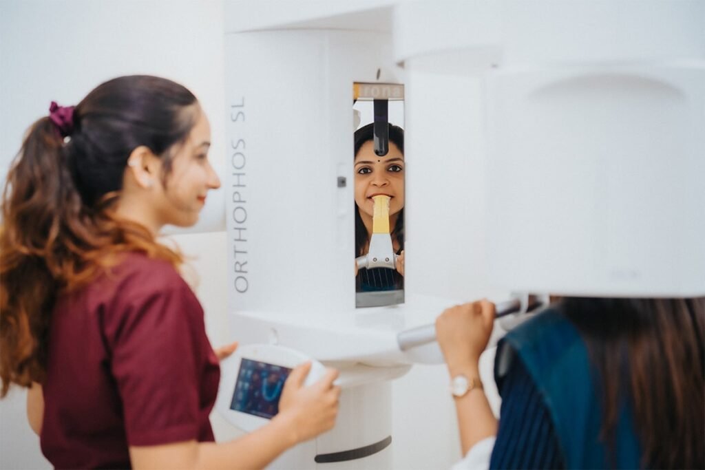
An OPG is a panoramic or a wide view x-ray of the lower one third of face, which displays all the teeth of the upper and lower jaw on a single film. It demonstrates the number, position and growth of all the teeth, including those that have not yet surfaced or erupted. Kamala Dental took a new step in upgrading by introducing the world’s best digital panoramic imaging system called Orthophos SL from Dentsply Sirona (USA).
There are clear advantages of panoramic images / OPG such as:

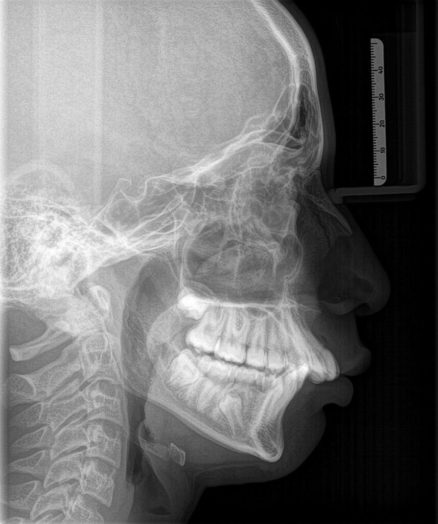
It is the best tool to assess the skeletal growth pattern of face for diagnosis in orthodontics and orthognathic surgeries. A Lateral Cephalogram is a lateral or side view radiograph of the face which demonstrates the bones and facial contours on a single radiograph. Various measurements can be made to determine the current and future relation of the upper and lower jaw (maxilla and mandible) and to assess the nature of a patient’s bite. Cephalograms can also help to predict mandibular growth in children by allowing accurate orthodontic measurements to be made and can help to determine the changes that have occurred during and after the course of treatment.
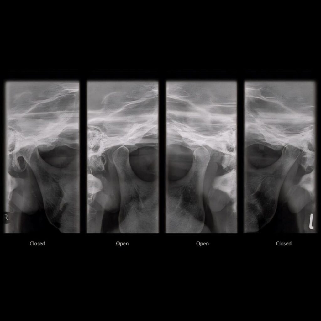
This imaging can diagnose disorders to treat them effectively. It offers both closed and open views of the joints. The axio-lateral temporomandibular view allows for visualization of various parts of the temporomandibular joint, such as the articular tubercle, mandibular condyle and fossa and it is useful to identify structural changes and/or any fractures as well as to assess joint spaces and excursion.
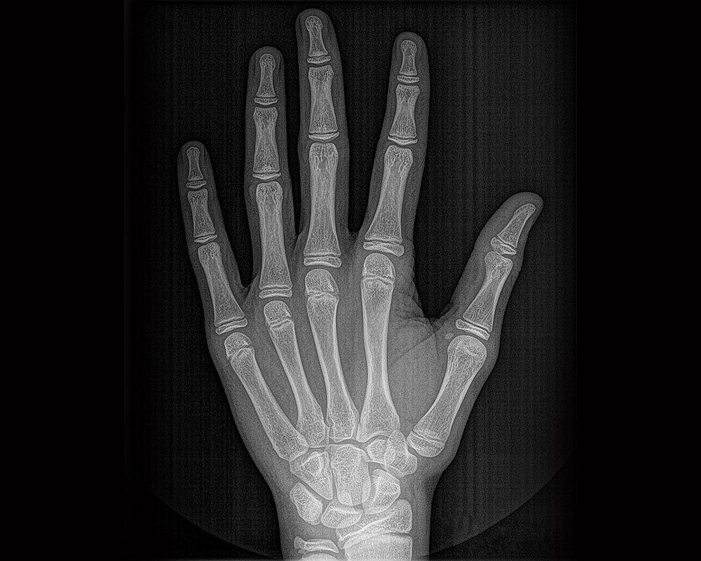
The hand and wrist radiograph help in estimating the skeletal age of bone for determining the physical maturation status of the child. It is used to determine parameters such as a patient’s skeletal age, the amount of growth left, to determine the type of orthodontic treatment or orthognathic correction required for the individual.