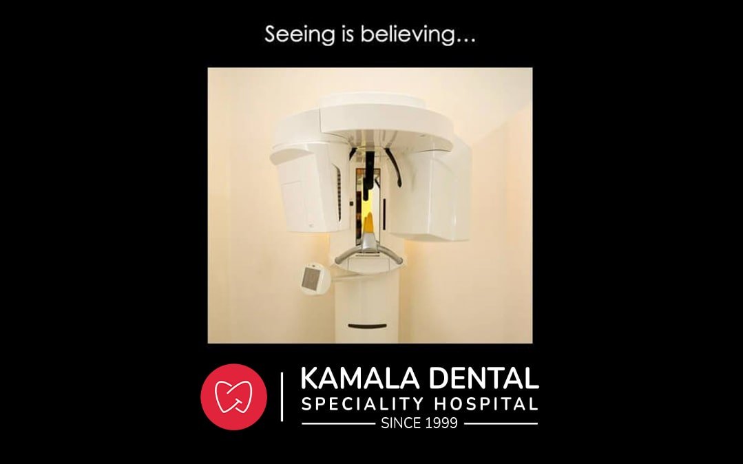
It is better to see and evaluate the teeth, bone and supporting structures in layers as it makes the surgical procedures more predictable and successful. Dental Cone Beam Computed Tomography (CBCT) is a special type of x-ray machine used in situations where regular dental x-rays are not sufficient. This scanner generates three dimensional (3-D) images of dental structures, soft tissues, nerve paths and bone in the craniofacial region in a single scan. Images obtained with cone beam CT allow for more precise treatment planning.
Metal objects, including jewellery, eyeglasses, dentures and hairpins, may affect the CT images and should be removed prior to your exam. You may also be asked to remove hearing aids and removable dental work.
It provides detailed images of the bone and is performed to evaluate diseases of the jaw, dentition, bony structures of the face, nasal cavity and sinuses. It has the advantage of lower radiation exposure.
Dental Cone Beam CT is commonly used for treatment planning of orthodontic issues. It is also useful for more complex cases that involve:
You will not experience any pain during a cone beam CT exam, and you will be able to return to your normal activities once the exam is complete. Your dentist, oral surgeon or radiologist will analyze the images.
Kamala Dental is equipped with latest CBCT machine of its kind – SIRONA Orthophos SL 3D with flat panel technology and utmost accuracy.